what type of chemical bond connects the complementary strands of a dna molecule to each other?
What do a human, a rose, and a bacterium take in common? Each of these things — along with every other organism on Earth — contains the molecular instructions for life, chosen dna or Deoxyribonucleic acid. Encoded within this DNA are the directions for traits every bit diverse as the color of a person'southward eyes, the olfactory property of a rose, and the style in which bacteria infect a lung cell.
DNA is found in nearly all living cells. However, its verbal location within a cell depends on whether that cell possesses a special membrane-jump organelle called a nucleus. Organisms composed of cells that contain nuclei are classified as eukaryotes, whereas organisms composed of cells that lack nuclei are classified as prokaryotes. In eukaryotes, Deoxyribonucleic acid is housed within the nucleus, but in prokaryotes, Deoxyribonucleic acid is located directly within the cellular cytoplasm, as there is no nucleus available.
But what, exactly, is DNA? In curt, Dna is a complex molecule that consists of many components, a portion of which are passed from parent organisms to their offspring during the process of reproduction. Although each organism's DNA is unique, all DNA is equanimous of the same nitrogen-based molecules. So how does DNA differ from organism to organism? It is simply the order in which these smaller molecules are arranged that differs amongst individuals. In turn, this pattern of organisation ultimately determines each organism's unique characteristics, thanks to another set of molecules that "read" the pattern and stimulate the chemical and concrete processes information technology calls for.
What components brand up Deoxyribonucleic acid?
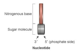
Figure 1: A unmarried nucleotide contains a nitrogenous base (ruddy), a deoxyribose saccharide molecule (gray), and a phosphate group attached to the 5' side of the sugar (indicated by light gray). Reverse to the v' side of the sugar molecule is the 3' side (night gray), which has a free hydroxyl group attached (not shown).
At the most bones level, all DNA is composed of a series of smaller molecules called nucleotides. In turn, each nucleotide is itself made up of three principal components: a nitrogen-containing region known every bit a nitrogenous base, a carbon-based carbohydrate molecule called deoxyribose, and a phosphorus-containing region known as a phosphate group attached to the carbohydrate molecule (Effigy 1). There are four different DNA nucleotides, each defined by a specific nitrogenous base: adenine (frequently abbreviated "A" in science writing), thymine (abbreviated "T"), guanine (abbreviated "G"), and cytosine (abbreviated "C") (Effigy 2).

Figure two: The four nitrogenous bases that compose DNA nucleotides are shown in bright colors: adenine (A, green), thymine (T, red), cytosine (C, orange), and guanine (Grand, blue).
Although nucleotides derive their names from the nitrogenous bases they contain, they owe much of their structure and bonding capabilities to their deoxyribose molecule. The cardinal portion of this molecule contains five carbon atoms arranged in the shape of a ring, and each carbon in the band is referred to by a number followed past the prime number symbol ('). Of these carbons, the 5' carbon atom is particularly notable, because it is the site at which the phosphate group is attached to the nucleotide. Accordingly, the expanse surrounding this carbon cantlet is known as the five' end of the nucleotide. Opposite the v' carbon, on the other side of the deoxyribose ring, is the three' carbon, which is not attached to a phosphate group. This portion of the nucleotide is typically referred to as the three' finish (Figure ane). When nucleotides join together in a serial, they course a structure known every bit a polynucleotide. At each point of juncture inside a polynucleotide, the 5' end of one nucleotide attaches to the iii' end of the adjacent nucleotide through a connectedness called a phosphodiester bond (Effigy 3). It is this alternating sugar-phosphate organisation that forms the "courage" of a DNA molecule.
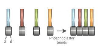
Figure 3: All polynucleotides contain an alternating sugar-phosphate courage. This courage is formed when the 3' stop (dark greyness) of i nucleotide attaches to the v' phosphate terminate (light greyness) of an adjacent nucleotide past way of a phosphodiester bond.
How is the DNA strand organized?
Although Deoxyribonucleic acid is ofttimes plant equally a unmarried-stranded polynucleotide, information technology assumes its most stable form when double stranded. Double-stranded DNA consists of 2 polynucleotides that are arranged such that the nitrogenous bases within one polynucleotide are attached to the nitrogenous bases within another polynucleotide past way of special chemic bonds called hydrogen bonds. This base-to-base of operations bonding is non random; rather, each A in one strand always pairs with a T in the other strand, and each C always pairs with a 1000. The double-stranded Dna that results from this blueprint of bonding looks much like a ladder with saccharide-phosphate side supports and base-pair rungs.
Annotation that because the two polynucleotides that brand upward double-stranded DNA are "upside downward" relative to each other, their sugar-phosphate ends are anti-parallel, or arranged in opposite orientations. This ways that one strand'south sugar-phosphate chain runs in the 5' to 3' direction, whereas the other'due south runs in the 3' to 5' management (Figure 4). It'south likewise disquisitional to empathise that the specific sequence of A, T, C, and G nucleotides within an organism'south Deoxyribonucleic acid is unique to that individual, and it is this sequence that controls not merely the operations within a particular cell, just within the organism every bit a whole.
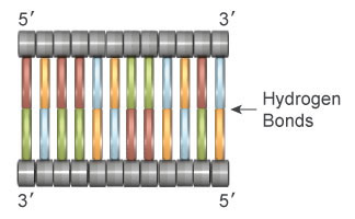
Figure 4: Double-stranded Dna consists of 2 polynucleotide chains whose nitrogenous bases are connected by hydrogen bonds. Within this arrangement, each strand mirrors the other as a upshot of the anti-parallel orientation of the carbohydrate-phosphate backbones, as well as the complementary nature of the A-T and C-1000 base pairing.
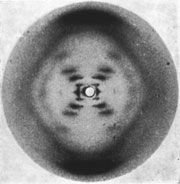
Figure five: Rosalind Franklin'due south X-ray diffraction image of DNA. Images like this 1 enabled the precise calculation of molecular distances within the double helix.
Beyond the ladder-like structure described above, another fundamental feature of double-stranded DNA is its unique 3-dimensional shape. The beginning photographic evidence of this shape was obtained in 1952, when scientist Rosalind Franklin used a process chosen 10-ray diffraction to capture images of Dna molecules (Effigy 5). Although the black lines in these photos await relatively sparse, Dr. Franklin interpreted them as representing distances between the nucleotides that were arranged in a spiral shape called a helix.
Effectually the same time, researchers James Watson and Francis Crick were pursuing a definitive model for the stable structure of Dna inside jail cell nuclei. Watson and Crick ultimately used Franklin's images, along with their ain evidence for the double-stranded nature of Dna, to contend that DNA really takes the form of a double helix, a ladder-like structure that is twisted along its entire length (Figure six). Franklin, Watson, and Crick all published manufactures describing their related findings in the aforementioned issue of Nature in 1953.
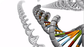
Effigy 6: The double helix looks like a twisted ladder.
How is DNA packaged inside cells?
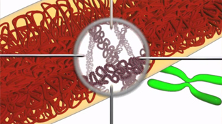
Figure 7: To better fit within the cell, long pieces of double-stranded Dna are tightly packed into structures called chromosomes.
Most cells are incredibly small. For example, i human being alone consists of approximately 100 trillion cells. Withal, if all of the Dna within simply one of these cells were bundled into a single straight slice, that Dna would be nearly 2 meters long! And then, how tin this much Dna exist made to fit within a cell? The reply to this question lies in the process known as Dna packaging, which is the phenomenon of plumbing fixtures DNA into dense compact forms (Figure vii).
During DNA packaging, long pieces of double-stranded Deoxyribonucleic acid are tightly looped, coiled, and folded so that they fit easily within the jail cell. Eukaryotes accomplish this feat by wrapping their Dna around special proteins called histones, thereby compacting information technology enough to fit inside the nucleus (Figure 8). Together, eukaryotic Deoxyribonucleic acid and the histone proteins that hold it together in a coiled class is called chromatin.
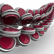
Figure 8: In eukaryotic chromatin, double-stranded DNA (grayness) is wrapped around histone proteins (red).
DNA tin can be farther compressed through a twisting process chosen supercoiling (Figure 9). Most prokaryotes lack histones, but they do have supercoiled forms of their Deoxyribonucleic acid held together by special proteins. In both eukaryotes and prokaryotes, this highly compacted DNA is and so bundled into structures called chromosomes. Chromosomes accept different shapes in different types of organisms. For instance, well-nigh prokaryotes have a single circular chromosome, whereas most eukaryotes have 1 or more than linear chromosomes, which oftentimes appear equally X-shaped structures . At different times during the life cycle of a prison cell, the DNA that makes up the cell's chromosomes can be tightly compacted into a structure that is visible under a microscope, or it can be more loosely distributed and resemble a pile of string.
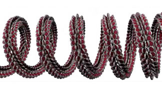
Effigy 9: Supercoiled eukaryotic DNA.
How exercise scientists visualize DNA?
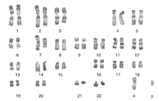
Figure ten: This karyotype depicts all 23 pairs of chromosomes in a human cell, including the sex activity-determining X and Y chromosomes that together make upward the 20-third fix (lower right).
It is impossible for researchers to see double-stranded Dna with the naked eye — unless, that is, they have a big amount of information technology. Modern laboratory technique permit scientists to extract DNA from tissue samples, thereby pooling together miniscule amounts of Dna from thousands of private cells. When this Dna is collected and purified, the consequence is a whitish, sticky substance that is somewhat translucent.
To actually visualize the double-helical structure of Dna, researchers require special imaging technology, such every bit the X-ray diffraction used by Rosalind Franklin. Yet, it is possible to see chromosomes with a standard light microscope, equally long as the chromosomes are in their most condensed form. To encounter chromosomes in this style, scientists must beginning use a chemical procedure that attaches the chromosomes to a drinking glass slide and stains or "paints" them. Staining makes the chromosomes easier to see under the microscope. In addition, the banding patterns that announced on private chromosomes equally a effect of the staining process are unique to each pair of chromosomes, so they allow researchers to distinguish different chromosomes from i another. And so, after a scientist has visualized all of the chromosomes within a cell and captured images of them, he or she tin can arrange these images to brand a blended picture chosen a karyotype (Figure 10).
Watch this video for a closer expect at the relationship betwixt chromosomes and the DNA double helix
Source: https://www.nature.com/scitable/topicpage/dna-is-a-structure-that-encodes-biological-6493050/
0 Response to "what type of chemical bond connects the complementary strands of a dna molecule to each other?"
Отправить комментарий